Ovarian reserve
- Pattern of menstrual cycle assessment (regular /irregular)
- Antral follicle count
- Serum FSH (menstrual Day 2-5)
- AMH – Produced by the ovary and does not fluctuate. At menopause can become undetectable.
Fundamental basis : women with good ovarian reserve have sufficient production of ovarian hormones from small follicles early in the menstrual cycle to maintain FSH at a low level. In contrast, women with a reduced pool of follicles and oocytes have insufficient production of ovarian hormones to provide normal inhibition of pituitary secretion of FSH, so FSH rises early in the cycle. The constant relationship between the number of follicles and circulating antimullerian hormone exists only after the age of 25 years. The recognition
that antim€ullerian hormone (AMH) is produced by preantral and small antral follicles with serum concentrations reflecting both the number of these small growing follicles and the primordial pool (1–3) now allows the opportunity for the study of aspects of these
earlier stages and factors influencing development, as well as the investigation of their relationship with fertility.
Perform basal cycle day 3 FSH
A normal result is not useful in predicting fertility, but a highly abnormal result ( FSH >20 mIU/mL) suggests that pregnancy will not occur with treatment involving the woman's own oocytes. A day 3 FSH concentration and a value less than 10 mIU/mL suggestive of adequate ovarian reserve, and levels of 10 to 15 mIU/ml borderline
Perform cycle day3 Estradiol : a cycle day 3 estradiol level, although there are conflicting data as to whether it is predictive of ovarian reserve and the response to ovarian stimulation (a value <80 pg/mL suggestive of adequate ovarian reserve
Antral follicle count (AFC) : Number of antral follicles (defined as follicles measuring 2 to 10 mm in diameter). On transvaginal ultrasound, a low AFC ranging from 4 to 10 antral follicles between days two and four of a regular menstrual cycle suggests poor ovarian reserve. Although AFC is a good predictor of ovarian reserve and response, it is less predictive of oocyte quality, the ability to conceive with IVF.
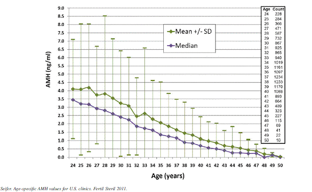
Anti-müllerian hormone (AMH) — Anti-müllerian hormone (AMH) is expressed by the small (<8 mm) preantral and early antral follicles. The AMH level reflects the size of the primordial follicle pool. In adult women, AMH levels gradually decline as the primordial follicle pool declines with age. AMH is undetectable at menopause.
serum AMH levels correlate strongly to antral follicle count and are more accurate than age and other conventional serum markers (FSH, E2, inhibin B) in predicting preovulatory oocyte supply in response to ovulation induction. Relative to the conventional ovarian serum makers, AMH appears to vary significantly less throughout the menstrual cycle.
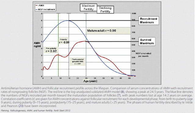
The circulating AMH also changes across the lifespan, with initial increases during childhood, then a distinct fluctuation around the time of puberty, followed by a secondary increase during the next decade to a maximum at around age 25 years. After the age 25, AMH undergoes a relentless decline to values below the levels of assay sensitivity between ages 40 and 45 years. This consistent decline in AMH from age 25 years is now firmly established.
After 25 years there is an uninterrupted and strong positive correlation betweenAMHand decreasing follicular recruitment as the pool of nongrowing follicles declines and eventually becomes exhausted at the menopause. It is this decline in follicular recruitment, albeit associated with
a maximal proportion of the available follicles achieving maturation, that results in a smaller number of follicles achieving the later stages of follicular development, which underlies the decline in AMH and explains the close relationship between AMH and egg yields seen in the IVF setting.
Antimullerian hormone is produced by growing follicles up to approximately 8 mm in diameter. Under normal circumstances much of the increase in circulating AMH arises after puberty.
PCOS and AMH : Women having PCO differ from those with normal ovaries by having slightly higher mean serum androgens or AMH levels. PCOs in normal women are not a morphological variant of normal ovaries but rather represent a functional entity that may be considered as a silent form of PCOS for which serum AMH could be the best marker. In conclusion, for the definition of PCOS, serum AMH appears as a sensitive and specific parameter that would probably be easier to reproduce from one to another centre than the follicle count, as the latter is highly dependent on the evolving quality of the machines and/or the operator skill. The threshold for AMH proposed to suggest the presence of Polycystic ovaries is 35 pmol/l (or 5 ng/ml).


No comments:
Post a Comment